


What are gallstones?
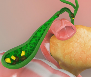
The gallbladder is a pear-shaped organ located on the right side of the abdomen, just below the liver. It stores bile fluid which is produced in the liver. Bile fluid contains water, proteins, fats, cholesterol, bile salts, and bile pigments, which aid in the digestion of food. The gall bladder releases bile fluid into the intestine through the common bile duct following the ingestion of food.
An imbalance in the components that form bile can lead to the formation of gallstones. Gallstones are small, hard deposits that form inside the gallbladder. The size of the gallstone can range from a small grain of sand to a large golf ball.
Gallstones are of two types:
- Cholesterol stones: These are yellowish-green in color and chiefly made up of hardened cholesterol.
- Pigment stones: These are dark and small, usually present in numbers and primarily made of bilirubin, a yellowish bile pigment. In some cases, a mix of both gallstone types can be seen.
Formation of Gallstones
Gallstones are usually composed of cholesterol or bile pigments. Lesser-known gallstones are made up of cholesterol and calcium salts.
Cholesterol stones or pigment stones may be formed due to:
- Excess secretion of bilirubin (an orange-yellow pigment) or cholesterol
- Low secretion of bile salts
- Presence of an infection
What is Choledocholithiasis?
The gallbladder is a small organ below the liver that stores a fluid called bile, necessary for fat digestion. The bile duct carries bile from the gallbladder to the small intestine. Choledocholithiasis is the presence of gallstones in the bile duct, causing obstruction. Gallstones can occur in the gallbladder and migrate to the bile duct, or they can form in the bile duct itself.
Symptoms of Choledocholithiasis
Symptoms include:
- Nausea and vomiting
- Fever
- Yellowing of the skin and eyes
- Severe abdominal pain
- Loss of appetite
- Clay-colored stools
Risks for Gallstones
Risk factors include:
- Obesity
- Liver cirrhosis
- Diabetes
- Low-fiber or high-fat diet
- Rapid weight loss diet
- Physically inactivity
- Pregnancy, from increased estrogen production
- Prolonged fasting
- Birth control pills or hormone therapy
- Hereditary disorders like sickle cell anemia
- Being of Native American or Mexican heritage
Diagnosis
Your doctor assesses your symptoms and performs a physical exam. Imaging and blood tests will be conducted.
Imaging tests include:
- Magnetic Resonance Cholangiopancreatography: This is an MRI of the pancreatic duct, bile duct, and gall bladder
- Transabdominal Ultrasound (TUS): Use of ultrasound to view the gallbladder or liver
- Endoscopic Retrograde Cholangiography (ERCP): A procedure to identify gallstones
- Abdominal CT-scan
Blood tests include:
- Liver function tests
- Complete blood count
- Bilirubin test
- Pancreatic enzymes test
Treatment of Choledocholithiasis
The treatment options include the following:
Surgery
Your doctor may suggest surgical removal of the gallbladder if conservative treatment options fail.
Laparoscopic/Robotic Cholecystectomy: It is the surgical removal of the gallbladder laparoscopically. A laparoscope is a thin fiber-optic device fitted with a camera and lens. Images from the camera are transmitted to a large monitor for your doctor to view the inside of your body.
The procedure involves the following steps:
- The surgery is performed under general anesthesia.
- You will lie on your back (supine position).
- Your surgeon makes small cuts in the upper abdomen.
- A laparoscope is inserted through one of the incisions.
- Special tools are used by your surgeon to isolate the gall bladder duct (cystic duct) and artery.
- The gallbladder is separated from the liver and carefully removed.
- The incision is closed and a bandage is applied.
Endoscopic Retrograde Cholangiopancreatography (ERCP)
This is a procedure that uses an endoscope to assist with removing stones from the bile duct. An endoscope is a long, flexible tube with a camera and light at one end that enables viewing of the internal organs. The procedure involves the following steps:
- An IV line may be used to provide a sedative.
- An endoscope is passed through your mouth into the duodenum (first part of the small intestine)
- A contrast dye is injected into the bile duct for visualization via x-ray.
- Special surgical instruments are guided through the endoscope.
- A small incision is made on the bile duct.
- Bile and pancreatic juices are drained out.
- A balloon or basket-type device may be used to collect the stones.


